
The University of Arizona is constantly making advances in cancer treatment. Two such examples in the area of radiotherapy are called intensity modulated radiation therapy (IMRT) and stereotactic radiosurgery/sterotactic radiotherapy.
Respiratory Gating
With CT information, physicians can see how much a tumor moves while the patient breathes. With this information and with the University's respiratory gating technology, radiation treatments can be monitored to more accurately focus the radiation on the tumor while giving less radiation to healthy tissue. The patient’s breathing is monitored, allowing physicians to know exactly where the tumor is during the breathing cycle. This information is then used to control the radiation treatment by turning the radiation on only when the area for treatment is within the planned radiation field. This enables the physicians to give more radiation accurately to the treatment area which increases the probability of controlling the cancer while reducing the risk of side effects.
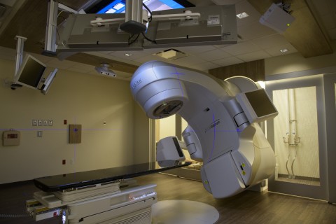
External Beam Therapy
In our facilities, radiation treatments are delivered using photon and electron beams produced by seven state-of-the art linear accelerators. The beams are shaped to conformally treat the tumor while minimizing the dose to healthy tissue and organs.
IMRT
IMRT is a sophisticated and effective radiation delivery system. It shapes the intensity of the radiation to deliver varying doses of radiation to small areas of tissue, conforming to the specific three-dimensional shape of almost any tumor. Doctors can deliver enough radiation to kill a tumor while sparing healthy surrounding tissue. Some IMRT techniques deliver a higher dose of radiation to the patient each day, potentially shortening the duration of the overall treatment and improving the potential for success. IMRT therapy may also lead to fewer side effects during treatment.
Brachytherapy
Brachytherapy is a method of using radiation to treat cancer by placing radioactive sources inside the tumor. It treats tumors from the inside out which results in more radiation to the tumor and less radiation to healthy tissue. Brachytherapy has been used for a variety of tumor types and sites.
In low dose rate (LDR) brachytherapy, small amounts of radiation are given over a long period of time. Radioactive sources, like cesium-137 or iridium-192, are placed in small tubes or needles and implanted into the tumor. The patient stays in the hospital for a few days or can be immediately discharged, depending on the isotope used. This form of therapy is especially useful for tumors where very high and localized radiation is needed to eliminate the cancer cells. LDR brachytherapy is often used for gynecological cancer and prostate cancer.
In high dose rate (HDR) brachytherapy, large amounts of radiation are given over a short period of time. Small tubes or needles are implanted into the tumor and then a small radioactive iridium source in inserted into the tubes or needles for a short period of time (5 minutes – 30 minutes). The source is then removed, the tubes or needles are removed, and the patient is discharged. HDR brachytherapy is often used for gynecological cancer, breast cancer, skin and prostate cancer.
Prostate Cancer Brachytherapy
Brachytherapy is a treatment for early stage prostate cancer, when the cancer is confined to the prostate gland. Prostate brachytherapy involves implanting tiny pellets containing radioactive iodine or palladium into the prostate tumor where the pellets give off radiation over time.
Using transrectal ultrasound (TRUS), an image of the prostate is visible on a monitor, and used for guidance when placing up to 100 seeds or more into the prostate. There is no need for a surgical incision. Instead, the radioactive seeds (which are smaller than a grain of rice) are contained in thin needles that are passed into the prostate gland through the skin between the scrotum and anus (Perineum). This is usually performed under either a spinal or general anesthetic. As the needles penetrate through the prostate they are seen on the monitor and can be accurately guided to their final position.
The radioactivity of the pellets slowly decays during the months after the operation, and there are few long-term risks associated with this treatment.
Facilities And Radiation Therapies
The University of Arizona's Department of Radiation Oncology is a well-equipped, state-of-the-art radiation therapy center. Capabilities include:
- High energy linear accelerators with MLC
- Low energy linear accelerators
- Stereotactic Radiosurgery/Stereotactic Radiotherapy
- CT simulator
- 3D treatment planning networked workstations
- Advanced IMRT and IGRT treatment planning
- Remote afterloading brachytherapy unit
- Low dose rate brachytherapy for prostate implants
- An array of dosimetric and quality assurance equipment